Joint and Muscle Flossing

Joint and Muscle Flossing

Have you heard of this relatively new treatment technique commonly called "flossing"? 

The rise in popularity of floss bands (or compression band therapy, CBT) can be largely due to interest from the Cross Fit world where it is a popular mobility tool for athletes pre competition and training. The idea and frequency of use in a clinical physiotherapy setting post injury or surgery is limited with a lack of good clinical trials currently available. Potentially, common injuries such as sprained ankles, torn muscles and post-operative surgery stiffness can benefit from such techniques.

The proposed benefits include:

  • increasing joint range of motion (ROM)
  • improved muscle mobility
  • decreasing pain levels
  • potentially speed up recovery through effect on myofascial release, occlusion and reactive hyperemia

Application

The practical use is generally limited to the joints and muslces of the periphery (the legs and arms for ease of application) though other areas have been "flossed". Stiff joints and/or tight muscles can be the target. Once shown this technique, floss bands can be self-administered as part of a warm up for training and competition to help tissue mobility. The process involves:

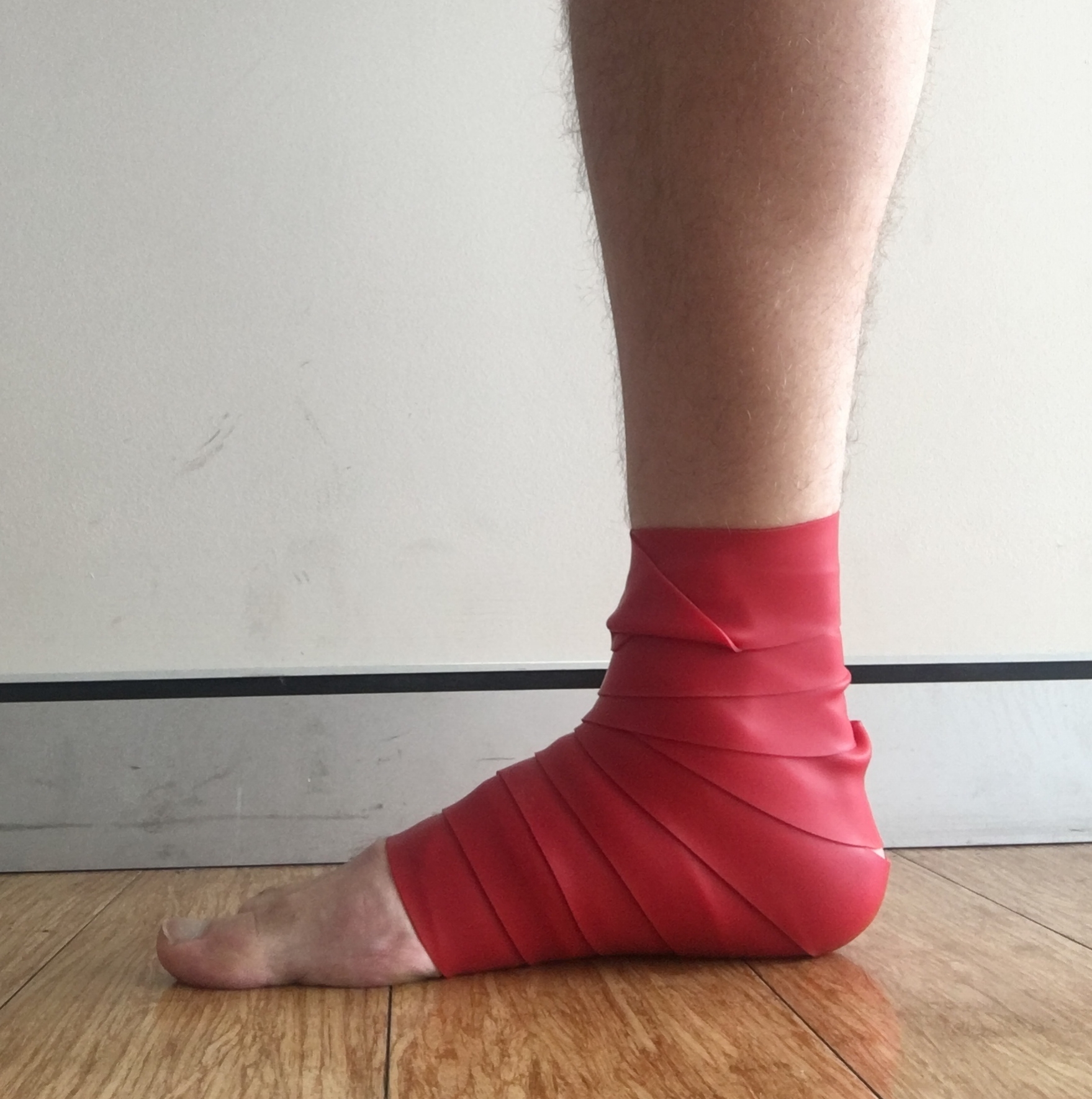
  1. Firmly overlap wrap the floss tape (2.5-7.5cm wide latex rubber band) around the limb (muscle or joint) from proximal to distal (although direction has not been scrutinized)
  2. Keep on for 2-4 minutes for treatment
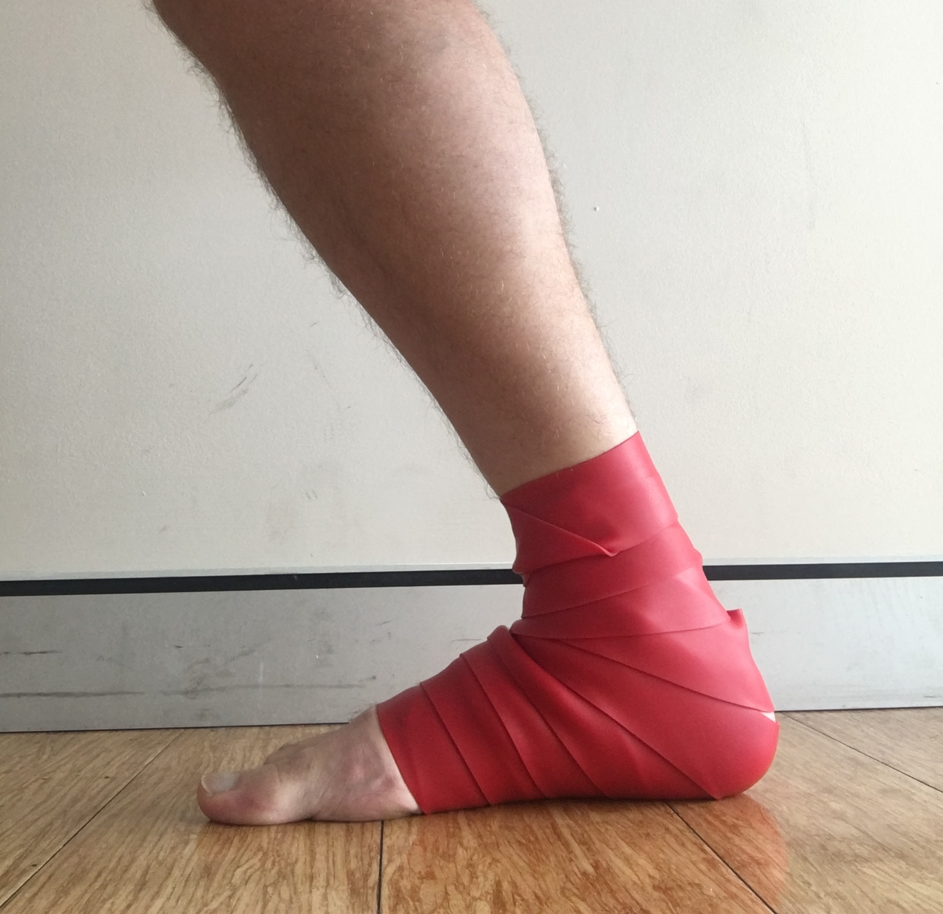
  3. The joint or muscle is moved through active or passive ROM (such as a squat/lunge/calf raise) (Figure 1)

An video example for floss application for a knee can be seen here

note: Neurovascular precautions should be observed during application to avoid numbness, pins and needles or excessive changes to blood flow

Figure 1. Ankle application and ROM

Research

Research into use of floss bands is limited, with scientific explanation of mechanisms and reliable research outcomes generally lacking. Bohlen et al. (2014) examined the effect of knee CBT on blood flow occlusion of the lower leg and found, while there was some improvement in strength, that reactive hyperaemic arterial blood flow showed no change. This would suggest that CBT does not alter blood flow significantly, though quality of this study was poor with a small sample tested (n=5). A search of several databases for clinical studies using floss bands or CBT revealed:

The primary theory of CBT is credited to fascial shearing or re-perfusion of blood to muscle (though as mentioned above the latter is less likely). The role of fascia is varied and can act to restrain motion, as a connective structure for skin or provide lubrication or gliding movement (Guimberteau, Delage, McGrouther, & Wong, 2010). The same group of researchers looked at fascial sliding in the hand during surgery with a visual description seen in Figure 2. Perceivably, with peripheral joints lacking bulky overlying tissue, this sliding of the superficial fascia is possible with CBT but yet to be researched. Another purported mechanism has suggested some psychological benefits following application of CBT to the shoulder (Kiefer et al., 2017).

fascia.png

Figure 2. Three-dimensional model of continuity between the skin and tendon provided by the sliding system (Guimberteau et al., 2010)

Conclusion?

At this stage any conclusions are merely anecdotal. Application of CBT/floss bands can potentially be a useful adjunct to current treatment techniques for acute joint sprain stiffness and restoring ROM. In my experience there seems to be a consistent effectiveness with ankle and knee ROM in the short term. Further review for the effectiveness with knee ROM post-surgery is warranted due to the increased presence of knee stiffness that is often seen. More studies are still needed though to get a better understanding of the proposed mechanisms of floss band treatment.

For more information or to give it a try ask one of your physiotherapists next time you see them!

Sanctband-2-Inch-Flossband.jpg

References

Bohlen, J., Arsenault, M., Deane, B., Miller, P., Guadagno, M., & Dobrosielski, D. A. (2014). Effects of applying floss bands on regional blood flow. International Journal of Exercise Science, Conference Proceedings, 9(2). 

Borda, J., & Selhorst, M. (2017). The use of compression tack and flossing along with lacrosse ball massage to treat chronic Achilles tendinopathy in an adolescent athlete: a case report. Journal of Manual & Manipulative Therapy, 25(1), 57-61. doi:10.1080/10669817.2016.1159403

Driller, M. W., & Overmayer, R. G. (2017). The effects of tissue flossing on ankle range of motion and jump performance. Physical Therapy in Sport, 25, 20-24. doi:10.1016/j.ptsp.2016.12.004

Guimberteau, J. C., Delage, J. P., McGrouther, D. A., & Wong, J. K. F. (2010). The microvacuolar system: how connective tissue sliding works. The Journal of hand surgery, European volume, 35(8), 614. doi:10.1177/1753193410374412

Kiefer, B. N., Lemarr, K. E., Enriquez, C. C., & Tivener, K. A. (2017). A Pilot Study: Psychological Effects of the Voodoo Floss Band on Glenohumeral Flexibility. International Journal of Athletic Therapy and Training, 1-16. doi:10.1123/ijatt.2016-0093
Knee pain whilst cycling: Does your bike fit?

Knee pain whilst cycling: Does your bike fit?

Do you get an increase in pain at the front of your knee after a ride? Has your bike ever been fitted to your bodies dimensions? This blog gets a bit into the science of how cycling related knee pain is caused and what you can do to prevent it.

The knee joint is one of the most common joints suffering from overuse injuries in cycling, accounting for more than 25% of injuries reported. The likelihood of knee pain (known as patellofemoral pain syndrome or PFPS) can be attributed generally to two factors:

  • Intrinsic factors:
    • changes in under or over-active musculature
    • mechanical variances such as increased Q angle
    • knee joint morphology
    • female gender
      • (Bailey, Maillardet, & Messenger, 2003; Dettori & Norvell, 2006; Ward, Terk, & Powers, 2005)
  • Extrinsic factors:
    • bike geometry and setup can increase patellofemoral joint (knee cap) stress resulting in knee pain
    • mileage and training load
      • (Asplund & St Pierre, 2004; Bini, Hume, & Croft, 2011)

The two primary causes of knee pain commonly seen in cycling by a physiotherapist are problems with muscle activation patterns and bike setup.

Muscle Activation Patterns

Muscle activity of the lower limb during pedaling has been studied, with the onset and offset of EMG activity in relation to crank position during pedaling shown in the figure below (figure 1). This shows the activation of the gluteals (GMax) and quadriceps (VM, VL, RF) (known as the power muscles) initiating before top dead centre (TDC) which are then followed by the hamstrings (SM, BF) and calf (GM, GL, Sol) (known as the coordination muscles). The power phase continues until bottom dead centre (BDC). Mechanical efficiency during pedaling is somewhat reliant on the activation and coordination of all active leg muscles and is not simply the result of any single muscle in particular. 

Figure 1. Duration of EMG during pedaling (Hug & Dorel, 2009)

The power muscles will generally increase in activation with increases in workload (i.e. go faster = work harder) whereas the co-ordination muscles change little but optimise a smooth transmission of power at TDC/BDC positions. Weakness of your glutes can lead to an increase in quadriceps workload and possibly increased kneecap stress. Riders with poor pedaling co-ordination are also likely to increase use of their power muscles at lower loads which can eventually lead to:

  • fatigue 
  • less efficiency
  • greater potential for stress throughout the kinetic chain(common in recreational cyclists)

So, if you are getting this kind of pain a few tips that are important to know include:

  1. Higher quadriceps forces --> higher knee cap compression forces --> greater risk of kneecap pain (PFPS) 
  2. Correct strengthening of power muscles can prevent this
  3. How to effectively time hamstring and calf muscle activity throughout TDC/BDC whilst pedaling to improve technique

Bike Setup

Several studies have looked at seat height as a causative factor in predisposing the knee to injury. The correct seat height will allow effective recruitment of your glutes and calf muscles as well as unloading the overstretched hamstring and neural structures. Common bike setup measures are shown in figure 2 below.

Figure 2. Common bike setup measurements (Wisbey-Roth & Visentini, 2016)

Measurement of correct seat height can be done with two common methods:

  1. Seat height (L5 measure in figure 2) as a percentage (usually 95-98%) of total leg length (measured as vertical distance of greater trochanter to floor) plus cleat sole height
    • This can involve a crank length (L6) adjustment
  2. Knee angle when the foot is at BDC and the cyclist is seated
    • For PFPS prevention the accepted angle is 25 degrees, slightly greater (30-35 degrees) for tendinopathies or ITB syndrome

A lower seat height (often seen in recreational cyclists) has been shown to increase contact forces at the kneecap joint and will often be the most likely cause of pain.   

Take Home Message

  • Seeing a sports physiotherapist can help identify if there are muscle weakness or activation problems and give you advice on how to correct these. This can lead to improved efficiency and reduction in pain. 
  • When treating cyclists with knee pain it is important to consider an optimal bike setup and riding position that is conducive to the ranges of the cyclist’s biomechanical limitations. This can not only minimise injury but also improve performance.
  • There are many elements to setting up your bike that can be altered (seat height is just one!). So if you suffer from pains in other parts of your body whilst riding (back, neck, shoulders etc) discuss this with your physio who can perform a bike-fit assessment or refer you to someone that can.


References

Asplund, C., & St Pierre, P. (2004). Knee pain and bicycling: fitting concepts for clinicians. The Physician and Sportsmedicine, 32(4), 23. 

Bailey, M. P., Maillardet, F. J., & Messenger, N. (2003). Kinematics of cycling in relation to anterior knee pain and patellar tendinitis. Journal of Sports Sciences, 21(8), 649. 

Bini, R., Hume, P., & Croft, J. (2011). Effects of Bicycle Saddle Height on Knee Injury Risk and Cycling Performance. Sports Medicine, 41(6), 463-476. 

Dettori, N. J., & Norvell, D. C. (2006). Non-Traumatic Bicycle Injuries. Sports Medicine, 36(1), 7-18. 

Hug, F., & Dorel, S. (2009). Electromyographic analysis of pedaling: A review. Journal of Electromyography and Kinesiology, 19(2), 182-198. 

Ward, S. R., Terk, M. R., & Powers, C. M. (2005). Influence of patella alta on knee extensor mechanics. Journal of Biomechanics, 38(12), 2415-2422. 

Wisbey-Roth, T., & Visentini, P. (2016). Optimising Biomechanics in Cycling (Course Notes). 

Low Back Pain in Golf

Comment

Low Back Pain in Golf

The who/how/why of low back pain in golf

Not an entirely uncommon occurrence following a round of golf for many of us, but this does not have to be the case.

Low back pain is generally considered to be the most common complaint by golfers. It has been reported that actual injuries to the low back occur in 11% of male golfers while the actual incidence of low back pain, in the same cohort, is 52% (Batt 1992). The Titleist Performance Institute (TPI) report that 28% of all players deal with low back pain after every round (TPI, 2015). Causes of low back injuries can vary and can include disc or facet irritation, stenosis with associated nerve impingement, degenerative joint disease and occasionally spondylosis (TPI, 2015).

"28% of all players deal with low back pain after every round"

The movements of the golf swing (figure 1) are generally referred to in phases of backswing, early and late downswing and early and late follow through (McHardy and Pollard 2005).  This large amplitude of movement involves rapid and forceful generation of power incorporating the hips/pelvis, trunk and upper limb transferring energy from the ground to the club resulting in considerable stress within the spine. 

These forces involve a downward compression, side to side (or lateral) bending and sliding (or back to front shearing) with the compressive loads at the 3rd and 4th lumbar vertebrae (L3/4) segment shown to peak at up to 8x body mass (Hosea et al 2010). Range of movement of the torso covers a large amplitude beginning in 30 degrees of forward flexion and by the end of the swing finishing in 30 degrees of extension (TPI, 2015). It is no wonder then that the discs that occupy the spaces between these vertebrae become vulnerable to damage.

bjsports-2005-November-39-11-799-F1.large.jpg

Figure 1. Phases of the golf swing. (A) Address position; (B) early back swing; (C) late back swing; (D) top of swing; (E) down swing; (F) acceleration; (G) early follow through; (H) late follow through. (McHardy and Pollard, 2005)

Lindsay and Horton (2002) looked at spine motion in elite golfers with and without low back pain and found that those WITH low back pain were inclined to have:

  • Increased spine forward bend when addressing the ball
  • Increased left side bend during the backswing (in right handed golfers)
  • Decreased trunk rotation in neutral resulting in a “supramaximal" rotation during the swing

They also showed that in golfers WITHOUT low back pain there was increased trunk flexion velocity during the downswing, implicit that increasing abdominal muscle activation, strength and force production during this movement may possibly be a protective mechanism (Lindsay and Horton 2002). 

Physiotherapy and Golf Injuries 

When having a physiotherapy assessment or screening, the questions usually asked should be sports specific questions relating to golf and golf pathology. These can include:

  • When does the player get pain (during or after a round of golf)?
  • What is the nature and intensity of the pain?
  • Past history of similar injuries?
  • What other injuries or aches and pain are present? (a painful Achillies tendon can speak volumes...)
  • Do they have lessons with a golf coach? If so, what are the common swing faults that have been identified? (biomechanical causes)
  • Volume of balls they hit per week? (overuse and load-monitoring)

When performing a physical assessment, areas that should be addressed first are often the dysfunctional non-painful movements. In other words, these are movements that the golfer is simply unable to do. This can be an inability to fully squat or having increased stiffness with thoracic rotation (click here for more info about the role the thorax has to play). They tend to be the biggest problems diagnostically and are generally the cause, not the site, of pain.

The physical assessment should also include a look at equipment and set up. Looking at the address position (golf stance) and posture can help determine likely causes and injury patterns and will usually identify when a poor movement pattern is present. What does their grip look like? What do their shoes look like? (including use of orthotics and wear patterns on the sole). Use of video analysis if possible can help identify common swing faults. These swing faults will also shed light on several injury inducing mechanics that can include: 

  • Increased Torque by not letting go of the lower body in the follow through and allowing the feet freedom to move
  • Excessive Right Side-bend at follow through will add to right side facet injuries and lateral rib stress injuries. This occurs by hanging back or having an increased closed club face
  • Early Extension of the hips can increase spine extension range and lead to facet compression
  • S-posture (likely associated with a lower cross syndrome) is usually a sign of tight hip flexors and inhibited abdominals and gluteals
  • Reverse spine angle (the spine should lean away from target not towards) creates unnecessary trunk movement and will increase the ROM (and subsequent velocity) of the spine during the swing that results in increased stress on facets and discs, more commonly in the right side of the lower back (in right handed golfers). Sway can exacerbate this fault

To discuss any of these issues or to have a golf specific screen get in touch to find out more.


References

Batt, M. E. (1992). A survey of golf injuries in amateur golfers. Br J Sports Med, 26(1), 63. doi:10.1136/bjsm.26.1.63

McHardy, A., & Pollard, H. (2005). Muscle activity during the golf swing. Br J Sports Med, 39(11), 799-804. doi:10.1136/bjsm.2005.020271

Hosea TM, Gatt CJ, Galli KM, et al. Biomechanical analysis of the golfer's back. In: Cochran, A. J. (2010). Science and Golf (Routledge Revivals). Florence: Taylor and Francis. Retrieved from http://ebookcentral.proquest.com/lib/latrobe/detail.action?docID=592967

Lindsay, D., & Horton, J. (2002). Comparison of spine motion in elite golfers with and without low back pain. J Sports Sci, 20(8), 599-605. 

TPI (2015). Course notes Level 2 Medical and online lecture material, Titleist Performance Institute.

Comment

Running with Hip Pain - Femoral Neck Stress Fractures

Running with Hip Pain - Femoral Neck Stress Fractures

Stress fractures commonly occur in sports where the movement demands of the sport produce repetitive loads on the body. Sports involving running, jumping and dancing place the individual at a higher risk of a bone stress injury. The incidence of stress fractures in the femur can range from 2.8% to 33% of all stress related injuries (both to the shaft and neck of femur). Management of these injuries requires an accurate diagnosis, often long periods of activity modification and rehabilitation (generally <4 months). The rehab program requires the close monitoring of a gradual and structured increase in load to allow the individual to return to full activity.

Stress fractures to the femoral neck are quite rare and are seen most commonly in long distance or marathon runners. While only accounting for a small percentage of all stress fractures they are considered to be quite serious given the length of time required for recovery and tendency to require surgical fixation if not addressed early. Differential diagnosis should therefore also consider other possible causes such as:

  • Bursitis
  • Tendonitis
  • Muscle strain or injury
  • Avascular necrosis
  • Slipped capital femoral epiphysis
  • Sacroiliac injury
  • Acetabular and pelvic fractures
  • Hip joint arthritis
  • Perthes disease
  • Femoroacetabular impingement (FAI)

What is a stress fracture?

A stress fracture can occur as the result of fatigue failure of bone where the amount of microscopic damage from repetitive load exceeds the bones ability to repair and remodel. The femoral neck is particularly vulnerable due to the fact that the majority of vertical compression forces load through the femoral head, falling medially to the shaft of the femur. This creates a compressive load medially and tension load laterally.

A stress reaction, or fracture, occurs when there is a failure of the bone to remodel adequately with the addition of repetitive sub-threshold stress. This ability of bone to adapt to pressure, or lack of it, is known as Wolff’s Law and refers to the bones anisotrophic ability to manage load along multiple axes. Cortical bone, of which the majority of the skeletal system is comprised, is heavy and has a slow metabolic turnover which can increased the likelihood of stress related injuries. Especially when there is inadequate recovery time after exercise and loading.

Mechanical load on the bone in the form of intrinsic loading (stress) or mechanical deformation (strain) is required for bone remodelling to occur. When a stress injury occurs the bone is unable to cope with the excess amount of stress or strain.

Factors to consider when determining the effect a load may have on the skeletal system would include:

  • Amount of load (volume, rate and frequency of load application)
  • Bone health and geometry (bone mineral density and anatomical cross sectional area can be used as measures of this)
  • Activity of surrounding muscle (where muscle strength and balance can have a protective effect on shearing loads) 

Load on a bone can be classified as compressive, tensile or shear. When a bone bends it will experience a compressive load on one side and a tensile load on the contrary side.  Usually a failure of bone integrity will occur on the tensile side. This can result in microscopic damage accumulating and fatigue failure of the bone occurring, described as “crack initiation” (Knaeding et al, 2005). Without adequate rest and recovery time, this process will continue towards a “crack propagation”  which results in a macroscopic failure of the bone.

Causes

Risk factors for developing stress fractures can broadly be categorised as intrinsic or extrinsic in nature.

Intrinsic factors:

  • Endocrine deficits (particularly in female athletes)
  • Bone geometry and density
  • Poor patterns of loading and pathomechanics
  • Inadequate nutritional profile
  • Vitamin D deficiency 
  • Overall physical fitness

Extrinsic factors:

  • Recent changes to training variables (such as increased frequency, intensity and duration)
  • Poor biomechanics
  • Poor set up 
  • Poor or incorrect equipment use
  • Footwear
  • Environmental considerations 

Diagnosis

Diagnosing a stress fractures of the femoral neck requires:

  • A thorough history exploring known risk factors:
    • Medications
    • Diet
    • Occupation
    • If female, menstrual history
    • Sudden increase in physical activity involving repetitive sub-maximal loads
  • Pain in the region of the groin or proximal femur (and occasionally asymptomatic knee referred pain)
  • Palpable pain of the hip joint and pain at the extreme end of range with passive hip joint movements
  • An active straight leg raise and log roll test may elicit groin pain
  • Functional tests such as single leg hopping have been shown to be positive in almost 70% of patients 
  • Imaging to confirm diagnosis:
    • Plain x-rays, while often negative in the early stages may show periosteal callous formation and intraosseous sclerosis. Plain x-ray sensitivity has been shown to range from 12-56% (Wright et al, 2016)
    • Gold standard is with MRI and/or bone scintigraphy. Sensitivity for bone scintigraphy is slightly less (50-97%) compared with MRI (68-99%) (Wright et al, 2016)
Table 1. Classification grade of stress fractures on MRI. &nbsp;*STIR – short tau inversion recovery

Table 1. Classification grade of stress fractures on MRI.  *STIR – short tau inversion recovery

Femoral neck stress fractures can be classified as compression or tension type:

  • Compression injuries generally occur at the inferomedial cortical bone of the femoral neck have better outcomes due to the reduced risk of displacement. These are usually managed conservatively with good outcomes
  • Tension injuries generally occur at the superolateral aspect of the femoral neck have been shown to have poorer outcomes and display greater rates of displacement. Due to this increase risk they must be diagnosed early in order to prevent poorer outcomes. These can often progress to a displaced fracture and inevitably end up with surgical internal fixation

Delayed diagnosis has been shown to be associated with poorer outcomes in athletes. A study of 23 athletes with femoral neck stress fractures with follow up over 6 years following injury noted the most significant aspect of management was a delayed diagnosis with 13 athletes requiring internal fixation. The average time to confirming the diagnosis was 14 weeks after initial onset of symptoms. The injuries requiring fixation were career ending for all elite athletes in the study (Johansson et al, 1990).

Management and Rehabilitation

Successful conservative management of femoral neck stress fractures requires:

  • early diagnosis
  • a graduated return-to-sport protocol -  guided by pain

The most important determinant to be made with femoral neck injuries is the site of stress. Lateral femoral neck injuries are considered a high-risk stress fracture compared with medial sided compression stress. As a result, a slower, more cautious conservative rehab approach should be taken with a longer recovery time for lateral sided compression injuries.

Management should be done in collaboration with a sports medicine physician who can address other possible intrinsic causes. Physiotherapy treatment is paramount as this will guide the athlete back to a sporting performance level. A guideline for time frames and loading is summarised in table 2.

Table 2. Time frames and guidelines for return to activity (Wang et al, 2015; &nbsp;Kaeding et al 2005)&nbsp;

Table 2. Time frames and guidelines for return to activity (Wang et al, 2015;  Kaeding et al 2005) 

Evidence for return to activity with femoral neck stress fractures is poor, lacking any substantial randomised control trials, and limited only to expert opinion based articles and case series (Wang et al, 2015). General consensus shows that a minimum period of 4-6 weeks of strict non-weight-bearing must be carried out until pain free. This can then be upgraded to partial weight-bearing, weight-bearing as tolerated and then followed by full weight-bearing. 

Continual and regular assessment is important to ensure the pain does not return and that suitable loading and subsequent healing has occurred. Follow up radiographs can be done to ensure adequate healing without progression of a fracture line. Non-impact exercises can then be gradually introduced followed by low impact and finally running exercise is allowed. Progression through such loads is only allowed in the absence of pain. Regression of load is needed if pain returns.

If you are a runner and have noticed a gradual increase in hip pain with running training, contact us at The Physio Lab for an opinion and discussion on what the possible causes might be and further management options to prevent a potentially serious injury from occurring. 


Kaeding, C.C., Yu, J.R., Wright, R., Amendola, A., Spindler, K.P. (2005). Management and return to play of stress fractures. Clinical Journal of Sports Medicine. 15(6), 442-7. doi: 10.1097/01.jsm.0000188207.62608.35

Wright, A.A., Hegedus, E.J., Lenchik, L., Kuhn, K.J., Santiago, L. and Smoliga, J.M. A (2016). Systematic Review with Evidence-Based Recommendations for Clinical Practice Diagnostic Accuracy of Various Imaging Modalities for Suspected Lower Extremity Stress Fractures. Am J Sports Med. 44(1), 255-263. doi: 10.1177/0363546515574066

Johansson, C., Ekenman, I., Tornkvist, H., & Eriksson, E. (1990). Stress fractures of the femoral neck in athletes: The consequence of a delay in diagnosis. Am J Sports Med, 18 (5), 524–528. doi:10.1177/036354659001800514

Wang, T., Matheson, G., and Safran, M.R. (2015) General Treatment Concepts for Stress Fractures. In Miller, T.L. and Kaeding, C.C. Stress Fractures in Athletes: Diagnosis and Management. Springer International Publishing, Switzerland doi: 978-3-319-09238-6

Breathing and Chronic Neck Pain

Breathing and Chronic Neck Pain

Stress can play a significant role in ischaemic muscle pain (a restriction in blood supply to muscle tissue) and/or neck pain. With this, the role of of breathing should be brought into the clinical equation. This is not only due to the proximity of the respiratory muscles to the thorax (rib cage) and neck, but also due to the connection breathing has with various emotional states in our lives. Think of how you hold your breath in reaction to shock or suspense or how your breathing is effected by anxiety or stress.

When I see patients that present with neck pain I would usually sub-classify them, in my own terms, as either ‘pathological necks’ (those with symptomatic pathology such as boney, muscular or neural) or ‘stress necks’. Not the most accurate descriptions, and often not so clean cut, but this basically enables me to identify that a 'stress neck' will probably not respond as well to traditional manual therapy techniques and is usually better off with modalities such as dry needling, generalised upper body exercises, breath work and general advice on stress management.

"Does a poor, weak, posture cause respiratory dysfunction?"

When managing neck pain that is the result of stress, a focus on breathing and breath work exercises can have a two-fold effect. Firstly, it can mobilise the ischaemic muscles that are, at least in part, the result of a static cervicothoracic spine. Many cervical muscles have their origins on the thorax and are associated with respiration. The effects of breathing can therefore result in improvements in neck and thorax posture. Secondly, there is a resultant stress reduction due to an increase in mindfulness surrounding the pattern and rate of breathing. Breathing techniques that slow down the out breath can help stimulate the parasympathetic part of the nervous system which can reduce stress levels in the body.

The respiratory system is involved in our bodies response to stress. Its role is to breathe faster to increase oxygen and blood circulation throughout the body. This system can be impaired by static postures - think of those hours sitting at the computer or in work meetings and the stiffness that occurs in the neck and upper back. The dysfunctional muscle patterns and poor breathing mechanics that result from these static postures will therefore be a contributory element in the way that the body reacts to stress and in its ability to bring the body back to a state of calm and rest. 

Dimitriadis, Kapreli, Strimpakos & Oldham (2016) recently reviewed the role a dysfunctional respiratory system can play in chronic neck pain. They extrapolated causality quite nicely in this model (figure 1) looking at the various mechanisms and manifestations of neck pain and how they relate to poor respiration. Interestingly they point out that drug use to manage pain can reduce respiratory drive, while on the other hand, noxious stimuli can increase respiration and alter rib cage mechanics. Both these changes lead to dysfunctional breathing patterns that can alter blood chemistry and result in hypocapnia (low carbon dioxide levels). This is often seen in patients with chronic neck pain. They do note that while changes are seen in respiratory function, they are not at a level that can be classified as pathological at this stage. More good quality trials are needed to better understand this relationship.

Figure 1


The same group of researchers (Dimitriadis, Kapreli, Strimpakos & Oldham, 2013) also showed that patients with chronic neck pain (noted as having pain for a period of at least 6 months with weekly episodes) compared to healthy matched controls, showed significant reductions in maximal inspiratory and expiratory pressures most likely related to poor global and local muscle systems. This may raise a chicken and the egg dilemma: Does a poor, weak, posture cause respiratory dysfunction or is it the other way around?


A simple method to teach diagphragmatic (or abdominal or belly) breathing is as follows:

  • Assume a comfortable position, usually lying flat on your back with your knees bent up, in a quiet and calm environment
  • Begin by relaxing your shoulders and arms
  • Place one hand on your chest and the other hand on your belly/naval region
  • Inhale slowly through your nose for 5-8 seconds
    • As you breathe in, your belly should rise and your lower ribs should expand outwards with minimal upper chest movement
  • Exhale slowly through your mouth relaxing your chest wall and abdomen, usually for the same duration (or slightly longer) than your breath in


The link between stress and neck pain is very important. Posture also plays a significant role in neck pain. This is where I think that bridging these two areas is necessary. Attention to the role of breathing mechanics and respiratory rate within the context of good alignment of the spine, is something were we, as physiotherapists and educators, can have a big impact on patients with stress related chronic neck pain.

For more information on physiotherapy management techniques of chronic neck pain please seek an opinion from your physio. 


Dimitriadis, Z., Kapreli, E., Strimpakos, N., and Oldham, J. (2016). Respiratory dysfunction in patients with chronic neck pain: What is the current evidence?, Journal of Bodywork and Movement Therapies, Article in press. Available online 8 February 2016. doi.org/10.1016/j.jbmt.2016.02.001.

Dimitriadis, Z., Kapreli, E., Strimpakos, N., and Oldham, J. (2013). Respiratory weakness in patients with chronic neck pain. Manual Therapy, 18(3), 248-253. doi.org/10.1016/j.math.2012.10.014

Running Pains

Running Pains

So you have finally decided to follow through with one of those new year resolutions. After dusting off the running shoes the kilometers are slowly starting to pile up and things are taking shape for the up coming running season. Only you notice a niggle, a slight ache creeping into your daily movements. Here we outline 5 areas where pain shouldn't be ignored. If this sounds like you, check in with your physio or sports doctor who can shed some light as to its possible cause and how to resolve it.


1. Iliotibial Band (ITB) pain

Probably one of the most common running injuries. Why should we worry about ITB, or lateral (outside) knee pain? Lots of runners get into trouble when they push on with this problem. The ITB is a thick band of fascia that extends from the outer side of the hip and pelvis to the outside of the knee acting as a stabiliser for knee mechanics during activities such as running. It extends from the tensor fascia late (TFL) and superior gluteus maximus muscles.  The quality of ITB pain is usually a relentlessly predictable pain on the outside of the knee which comes on every time you reach 'x' kilometers. It is pointless attempting to push on through the pain (many try and fail) and why should you when there is plenty you can do about it.

Using a foam roller, the go to treatment for this condition, is rarely enough. Most will often only roll the band itself without addressing the associated TFL or glute max tightness. It usually is a little more multifaceted where you may need gait retraining to change some non-optimal biomechanics. For example you might have:

  • a stiff hip
  • poorly utilised glutes
  • reduced thoracic control and movement
  • inappropriate foot wear

Many people have a gait pattern that is too narrow and their feet aren't widely spaced enough, this leads them to scissor across at the mid line during running resulting in a tightening of the this tissue. If there is a focal ITB problem (swelling or thickening), you might end up needing a ‘rescue injection’ just before that marathon (which still might not guarantee you’ll get around comfortably). This will require a visit to a qualified sports physician. The moral of the story is, get help early with an experienced running physiotherapist, who can help you make these changes.

2. Hip and Groin Pain

There is really no such thing as a “Groin Strain” and despite what we might be told, its actually not that common to have a true hip flexor problem. Groin pain often results from the many structures around the hip and pelvis. Whilst you might be tight in your hip flexors, more often pain at the front of the hip is due to hip impingement (a.k.a. ‘FAI’), overload of the pubic bone area or problems at the back of the pelvis at the sacroilliac joint (SIJ).

Probably the most serious cause of groin pain in runners is a stress fracture involving the neck of the femur. It can have disastrous consequences if not managed properly. You might have the beginnings of a stress fracture if:

  • You can feel pain in the groin every time your foot strikes the ground
  • You have ‘random’ pain which ‘comes and goes’ and seems to move around
  • If you are “aware” of your niggle at night time.

 It’s really important that you get checked out early, and you may need an MRI scan to do this. X-rays are not sufficient to detect stress fractures. 

3. Foot pain

Metatarsal stress fractures are common in runners who present with foot pain, usually with a history of a gradual onset of symptoms that are slow to settle. Pain in the ball area of the foot might also be the result of a neuroma or sesamoiditis (inflammation of two pea sized bones under your big toe).

A neuroma feels like a lancing or knife like pain, between the heads of the metatarsal bones. It occurs when a nerve gets pinched, often between the 3rd and 4th toes, and results in a local swelling and inflammation around the nerve. It is often more common particularly if the arch across the front of your foot is flattening out. Neuromas can be made better with certain taping techniques, some appropriate orthotics from a podiatrist, alterations and gait pattern re-education. Some may even benefit from a cortisone steroid injection by a Sports Physician.

Sesamoiditis feels like an intense pain under the ball of your big toe felt at push off or in pivot sports such as golf. It is easily managed with so simple orthotics or padding to offload the effected area in combination with some load management strategies.

Running with a foot stress fracture (seen mostly in the navicular or head of the 5th toe) can grind you to a halt requiring an extended period of rest and possibly surgery. These injuries often require a period of rest (sometimes in a boot) to allow the bones to settle. Appropriate imaging is needed to get an accurate diagnosis to plan bow best to manage them. 

Always seek a proper diagnosis with these symptoms. All may not be lost and seeking advice early is the best bet.

4. Heel pain

Sometimes plantar fasciitis isn’t plantar fasciitis! Sometimes pain in the area of your heel can be a calcaneal stress fracture or nerve irritation. Plantar fasciitis needs a proper biomechanical work up (usually from head to toe as the problem does not start in the foot but merely finishes there). Questions that should be asked include:

  • Do you have a stiff ankle or foot, which reduces its ability to re-distribute ground reaction forces, which overloads the plantar fascia? 
  • Are your foot intrinsic muscles working correctly?
  • Do you have a poorly functioning thoracic rotation movement pattern or reduced hip mobility?
  • Do you have weak soleus calf muscles, or a tight calf complex?
  • Maybe you simply need some different footwear or orthotics?

Shockwave therapy has been shown to be helpful in some resistant/specific cases of plantar fasciitis. Great Physiotherapy work will help you to resolve this more swiftly than you imagine, so don’t push on through the pain. Identifying where your body is failing to load correctly is often the most effective management strategy in the long term.

5. Shin splints

Particularly if these are severe you might actually be running the risk of a tibial stress fracture rather than just overload of the junction between the soft tissue and the bone. It is not uncommon to see patients post-marathon who actually have run (in agony) with a tibial stress fracture, whilst believing that they had simply a ‘bad case’ of shin splints. DON’T be tempted to run through this.  It can end in a very, very length rehabilitation process (or even surgery) to fix the bone. Addressing poor foot intrinsic muscle function a control is a great place to start. Stand up and try to bend and flex your big toe in isolation from your other toes while standing... having trouble getting the message across? 

Get proper physio advice for this and other intrinsic exercises is a good place to start managing painful shins. They can assess the injury to determine how to best manage such problems. If there is any doubt, they can then recommend you seek the specialist attention of a Sports Physician early on who can get appropriate imaging and blood tests to determine if there is an underlying cause.

All of these conditions, if given early attention, can typically be resolved swiftly and conclusively. So don’t grit your teeth and jog-on with pain. Get it sorted!


Note: This article was adapted with approval from a previous version written by Dr Cath Spencer-Smith - an Exercise and Sports Medicine Doctor based in London UK (more about Cath at http://www.sportdoclondon.co.uk/ )