Managing a Hamstring Tear

Comment

Managing a Hamstring Tear

One of the most common muscle tears an athlete will have, is a tear of the hamstring muscles. It can put an athlete out for the season if it is not managed correctly. Hamstring tears can originate in various parts of the muscle (the origin or insertion, muscle tendon or muscle belly) and differ in severity of grade. 

It is usually the result of over exerting the muscle during a sprinting style activity, although a fall, slip or sudden movement may also result in the injury occurring. An early, accurate, diagnosis by a physiotherapist and appropriate treatment will provide the best chance of returning safely to sport in a timely manner.

Anatomy 

The hamstrings are a group of 3 muscles found in the posterior (rear) region of the thigh, extending from the buttocks to either side of the knee. They are some of the longest muscles in the body and are diarthoidial in nature, which means they extend across two joints (the hip and the knee). This last point is important when examining the action of the muscles, as both joints are implicated when an injury occurs. 

The hamstrings can be divided into 2 groups; the medial (inside) and lateral (outside) groups. The medial group is made up of the semimembranosus and semitendinosis. The lateral group consists of the biceps femoris (long and short head). The primary function of the hamstrings is to flex (bend) the knee. It also has lesser functions to extend the hip and control rotary forces at the knee at end of range. They are primarily innervated by the tibial nerve. 

The attachments of the hamstrings all originate at the ischial tuberosity (the short head of the biceps femoris originates at the posterolateral femur). The semitendinosisinsertion fans out in the shape of a ducks foot (pes anserine) and joins with the tendons of Sartorius and Gracilis as it attaches just above the tibial insertion of the medical collateral ligament of the knee. The semimembranosus is attached to the posteromedial tibia. The lateral insertion is less complicated although it incorporates its attachment at the head of the fibula with the arcuate complex at the posterolateral corner of the knee.  

(NB: These variations in attachments are important as often an injury to the hamstring can implicate the medical collateral ligament or the posterolateral corner of the knee) 

Causes 

There are many different factors that can predispose an athlete to a tear:

  • Poor flexibility of the muscles  
  • Reduced or side-to-side differences in strength of hamstrings   
  • Increased neural tightness, often arising from the lower back 
  • Inadequate warm up 
  • Fatigue 
  • Poor biomechanics or running pattern
  • Return to activity prematurely following previous injury 

Grade of Tear

Grade 1

  • Usually a mild strain where cramping or tightness may be felt. Often there is some pain when the muscle is stretched or palpated. 
  • Recovery: 5 days-4 weeks (depending on recurrence and ability to perform a sports specific program pain free). 

Grade 2 

  • This grade usually is associated with more marked, instant, pain causing immediate cessation of activity. Athletes often describe a “ping” feeling, like a rubber band has snapped. This is confirmed again with palpation and stretch/contraction pain. There may be signs of swelling and often delayed bruising in and around the area. 
  • Recovery: 4-6 weeks

Grade 3 

  • This is a complete rupture and is quite rare. Usually associated with a slip or fall into a splits position or jerking movement (often with water skiing). There will be immediate loss of strength. Pain is usually present, but not always. There will be a palpable area of swelling and a depression where there is a tear.  
  • Recovery: Complete tears require surgical reattachment followed by a 3-6 month specific rehabilitation program 

Physiotherapy and Rehabilitation 

Optimal recovery from an injury to the hamstrings requires accurate diagnosis and grading. The length of time from injury is important in determining a suitable program. An individualised rehabilitation program should follow a basic timeline of:

  • acute/subacute phase
  • repair/remodelling phase
  • functional/return to activity phase  

Timelines for the use of Non-Steroidal Anti-Inflammatory Drugs (NSAIDs) has been debated with some professionals opting for immediate use (to prevent adverse effects from the injury that may interfere with tissue remodelling) and others suggesting a delay of 2-4 days post injury (to prevent interference with the inflammatory response and the laying down of new muscle fibers). It is best to discuss this with your treating physio or doctor. 

Acute/subacute Phase (1-14 days) 

The goals of this phase include reduction of pain and swelling and restoration of normal range of motion (ROM). This initial treatment follows the general principles of RICE (rest, ice, compression, elevation) and may involve the use of aides with more severe injuries. Once pain is reduced, gentle soft tissue massage and ROM exercises can begin, progressing from passive to active-assisted to active movements.  

Eventually flexibility and strengthening exercises can be started, commencing with closed chain. Cardiovascular exercise can commence, such as cycling, swimming and light jogging. A graduated running program can be commenced once pain free and is progressed accordingly (see running program). 

It is important to perform adequate strengthening, flexibility and balance exercises as advised by your physiotherapist. Returning to activity too early can pre-dispose to re-injury. 

Repair/remodeling Phase (1-6 weeks) 

During this phase strengthening can be progress to open chain concentric exercises, initially with low weight and reps and slowly increased. This can then be progressed to eccentric exercises but should be done carefully at first so as to avoid over exertion and lumbo-pelvic compensations. Strength should be progressed to reflect similarity with the unaffected leg. At the same time flexibility and balance (proprioception) should be worked on and progressions through the running program. 

Other modalities such as ice/heat, dry needling, taping and joint mobilisation may also be used throughout this phase. 

Functional/return to activity Phase (2 weeks-4 months) 

By this phase there should be no pain, normal gait pattern, good strength and ROM. Functional, sport specific exercises can gradually be incorporated into the program after careful discussion with your physiotherapist.

This is where the graduated running program is very useful as is provides a suitable progression of intensity and volume of activity. Plyometric and dynamic exercises can also be added to improve speed and power. Extrinsic variables such as use of equipment and balls can also be introduced in this phase. The patient should be reminded of the importance of a warm up and should continue doing flexibility exercises. 

Surgical Repair 

The need for surgical intervention is rare but generally required for grade 3 complete ruptures when the tendon has come off the bone. The time frame from injury to surgery is variable with good results in both acute and chronic cases (although acute intervention is preferred).


References
Warren P, Gabbe BJ, Schneider-Kolsky M, et al. Clinical predictors of time to return to competition and of recurrence following hamstring strain in elite Australian footballers. Br J Sports Med. Aug 14 2008;[Medline]. 

Agre JC. Hamstring injuries. Proposed aetiological factors, prevention, and treatment. Sports Med. Jan-Feb 1985;2(1):21-33. [Medline]. 

Clanton TO, Coupe KJ. Hamstring strains in athletes: diagnosis and treatment. J Am Acad Orthop Surg. Jul-Aug 1998;6(4):237-48. [Medline]. 

Sallay PI, Ballard G, Hamersly S, et al. Subjective and functional outcomes following surgical repair of complete ruptures of the proximal hamstring complex. Orthopedics. Nov 2008;31(11):[Medline]. 


Comment

THE THORAX IN GOLF

Comment

THE THORAX IN GOLF

It is an all too common story in golf. An acute or overuse injury to the lumbar spine will result in a change in swing mechanics, poor shot selection and a few lost balls. This is due to pain it causes or physical impingement that results, or both.  So after repeated treatments with your local physiotherapist to manage the low back pain only give temporary relief at best. Then some x-ray or MRI images are sort after which pain the picture of a degenerative back with possible disc damage. Next stop is the orthopaedic surgeon for surgery to fix the picture. The problem is the problem never really goes away. What if the lumbar spine is not the problem in the first case?

How many of you have walked into a clinic expecting to be treated for your lower back only to find yourself having some manual therapy done on your thorax (mid back)? This combined with some exercises that address the mobility and control of your mid back are what you then work on. Don’t be too surprised to find the lower back pain will often then lessen or, better still, disappear.

Does a pain-free thorax then equate to ideal mechanics in your spine?

The Thorax

The thorax is inclusive of the thoracic spine, the ribcage, chest plate and internal organs, forming 20% of our total body length. Contrary to popular belief, the thoracic spine works in combination with the ribs attaching to each vertebrae, forming a ring. It is the function of these rings in relation to each other (translation and rotation) that gives the thorax its mobility. For further information about how these thoracic rings work click here.

The thorax often gets under or misreported as lower back pain in a lot of clinical data. It is in the thorax predominantly that most of the rotation of the spine occurs. Compensatory rotation of the lumbar spine, especially when combined with flexion (bending) such as during a golf swing, can make the lumbar discs increasingly vulnerable to injury. The lack of thoracic rotation is seldom due to stiffness but instead a lack of adequate neuromuscular control of the joints in the thorax.

Now consider the number of muscles that attach from your thorax to your pelvis, head/neck and shoulders, all of which are pivotal in transitioning force generation from the pelvis to the upper limb in the golf swing. Lower thorax issues will most often impair and alter the functional movements of the pelvis resulting in loss of stability, strength and movement. Similarly, a poor upper thorax will alter the ability to lift and rotate the shoulders, arms and head/neck.
 

IMG_0127.jpg

The Body-Swing Connection 

The sequence of body segments during the downswing (in a right hander) follows first the pelvis translating and rotating to the left, then rotation of the thorax, then arms and club back toward the ball. Dysfunction in the thorax will prevent the optimal transition of segmental movement from your lower body into your upper body and club. This can result in any number of the compensatory swing faults commonly seen. This impaired swing is also less effective (resulting in shorter and less accurate shots) and may also lead to further injury and overuse of other body segments.

C09F1C4E-071D-494C-AC0E-EBD356F31AD1.JPG

A simple test to determine whether or not your thorax is affected and contributing to the dysfunction is to:

  1. Sit on the edge of a chair, fold your arms across your chest

  2. Turn as far as you can with your upper body, without moving your backside off the chair

  3. Compare this from left to right, can you turn 90 degrees to both sides?

Questions to ask:

  • Is there one side tighter than the other?

  • Is there pain? Where is the pain?

  • Are you actually moving your thorax versus turning your neck, side bending your trunk or pulling your shoulders around?

Treating a Dysfunctional Thorax

Treating the thorax may involve manual therapy by a physiotherapist to release fixated joints or normalise over/under active muscles that are preventing optimal movement. The body is an incredibly interconnected unit and more than just the sum of all its parts. Often there may only be one or two primary areas creating this suboptimal movement, which is generally referred to as a 'driver' as it is the lead cause of the problem (or primary and secondary drivers where there is more than one).

Once the driver has been identified (this may not necessarily be the thorax but can be elsewhere in the body) and normal motion is restored to that area, it is important to then retrain the brain to make effective use of the increased range of movement and to regain neuromuscular control of that area. Knowing where and what your driver is can help you address the problem with greater efficiency. This is where communication between your golf coach, physiotherapist and fitness trainer is important.

"Knowing where and what your driver is can help you address the problem with greater efficiency"

Motor control exercises to correct the primary driver and facilitate new muscle patterns are important to prevent unwanted muscle tone and postures from returning following treatment. Ever walked out of the therapist treatment room feeling great only for this to disappear 2 days later? Exercises to retrain and strengthen the area of treatment play a vital role and can include simple movements where specific segmental muscles are cued with appropriate feedback (often with tape, biofeedback or use of mirrors).

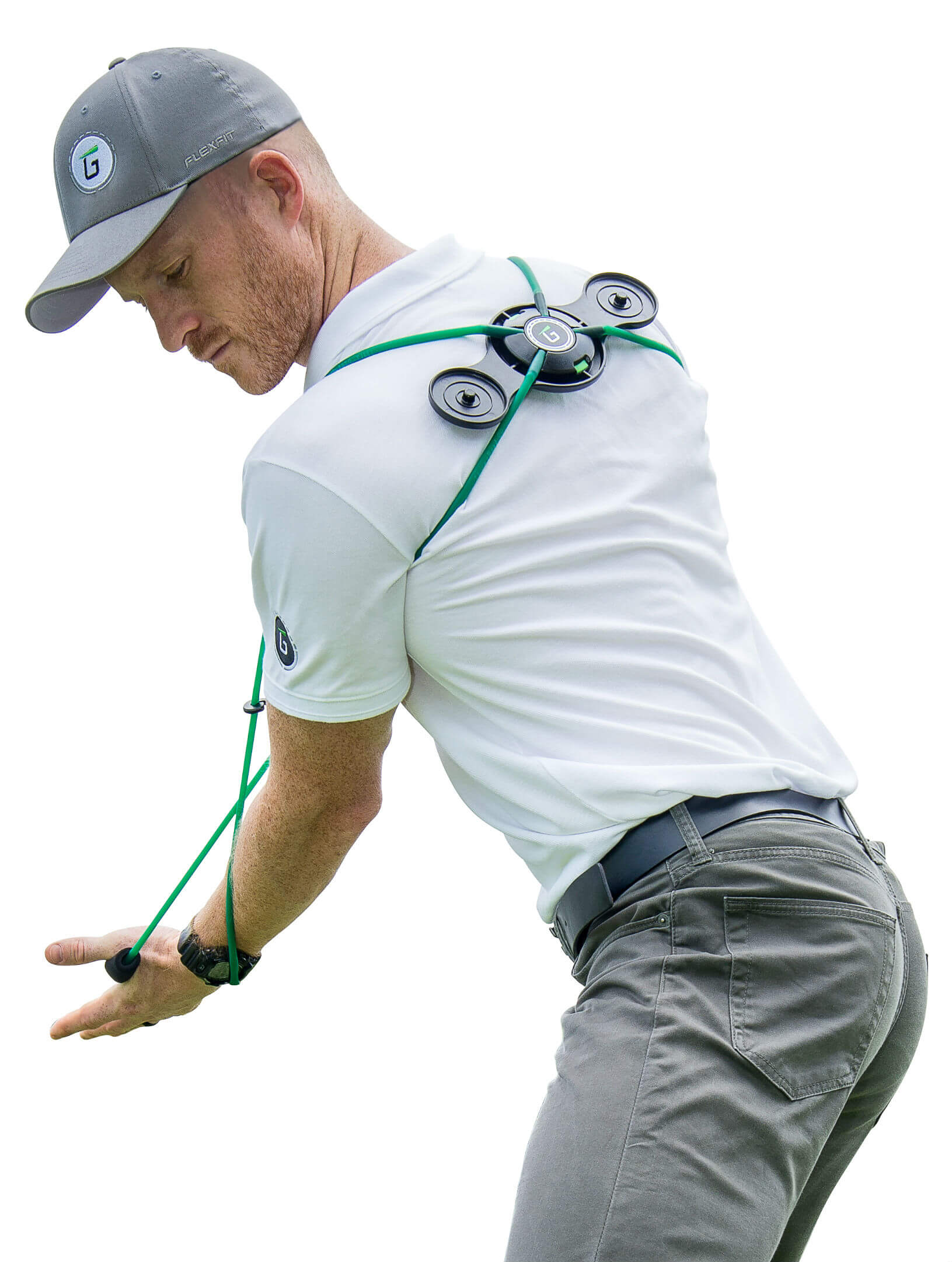
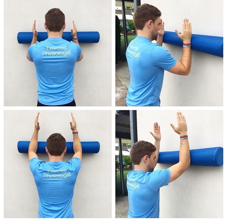
Gaining better activation awareness and strengthening of the serratus anterior muscle can obtained with exercises such are wall slides (as seen below) and with the use of resistance bands. Devices such as the TPro (from Gravity Fit) can aide and improve in feedback of the thoracic control muscles during various rotational movements that mimic the golf swing. It can also then be taken on range to incorporate this training into sport specific practice once the correct skill is obtained.

Appropriate thoracic strength and power exercises, and subsequent golf specific drills, can be added to increase performance measures. It is important to maintain the correct cueing pattern throughout, until such a time that the correct movement becomes automatic. Understanding where your driver can simplify this process and is key to effective long term management of such musculoskeletal conditions.

Treating the thorax may not fix your golf swing but it will enhance your movement pattern and ability to sequence body parts. This then decreases the chance of acutely injuring your back on the course or creating an overuse condition. Once you can optimise the fundamental movement components of a swing progress with your teaching professionals can help lead to improvements on the course.

For further information about a back pain in golf, or to have a golf screen, click here. For a physiotherapy appointment click here to book online.

Comment