What the Evidence Says (Lead vs Trail Leg)
Returning to golf after anterior cruciate ligament reconstruction (ACLR) isn’t just about healing — it’s about restoring mechanics, strength, and confidence to safely tolerate the unique demands of the golf swing. Golfers need to understand how lead and trail legs differ biomechanically during the swing, as this impacts rehabilitation priorities and safe return to play.
ACL Recovery & Return to Sport (RTS): What the Research Shows
RTS Outcomes Are Multifactorial
Return to sport following ACLR depends on more than the surgical procedure itself. Strength, movement mechanics, functional performance, and psychological readiness all contribute to successful RTS. A recent scoping review emphasises the variability in RTS criteria and highlights the importance of objective assessments, such as quadriceps and hamstring strength, as well as hop tests, typically requiring ≥90% limb symmetry before considering full return to play (Wright et al., 2025).
While ACL reconstruction is common for athletes aiming to return to sport, some meta-analyses show similar return-to-sport rates between reconstruction and structured rehabilitation alone, especially when high-quality rehab is performed. This highlights the critical role of functional recovery over surgery per se.
Functional Performance Predicts Long-Term Outcomes
Performance testing around 6 months post-ACLR can predict long-term return to pre-injury activity at 12–24 months. Quadriceps strength, single-leg hop distance, and other functional tests provide insight into knee stability, limb symmetry, and readiness for sport-specific demands, including golf (Nawasreh et al., 2018). Relying solely on time since surgery is insufficient and use of functional benchmarks are critical.
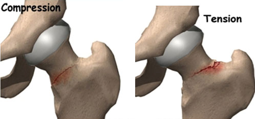
Persistent Biomechanical Deficits
Even athletes cleared for sport often demonstrate kinematic and kinetic asymmetries, which can increase reinjury risk. These deficits are frequently observed in knee rotation, frontal-plane control, and eccentric load absorption. This underlines the importance of comprehensive, objective RTS assessments rather than clearance based on surgical milestones alone (Alarifi et al., 2025).
Even basic movement patterns such as gait may show ongoing interlimb biomechanical asymmetries up to 6 months post-ACLR, suggesting that restoration of knee mechanics often lags strength recovery
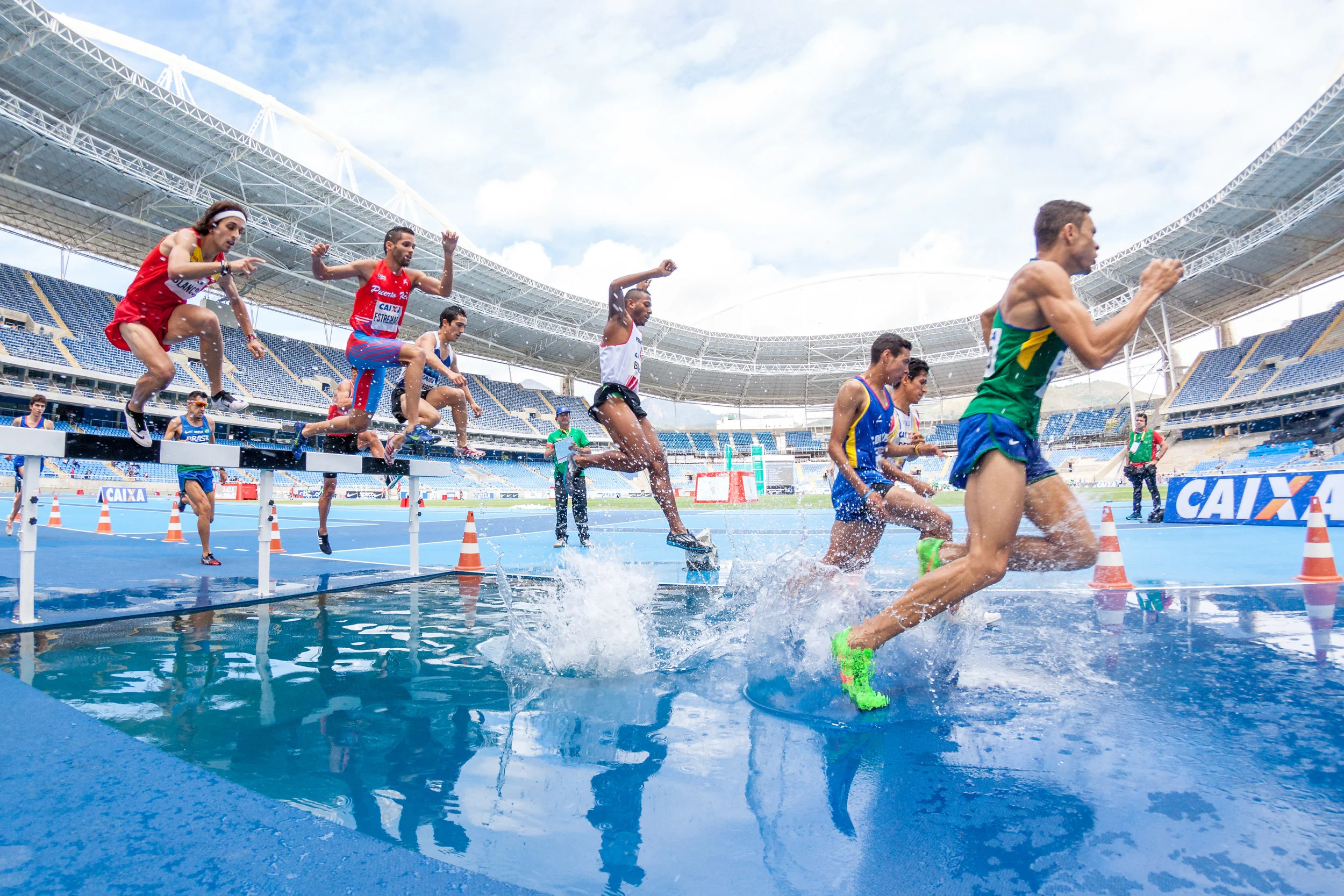
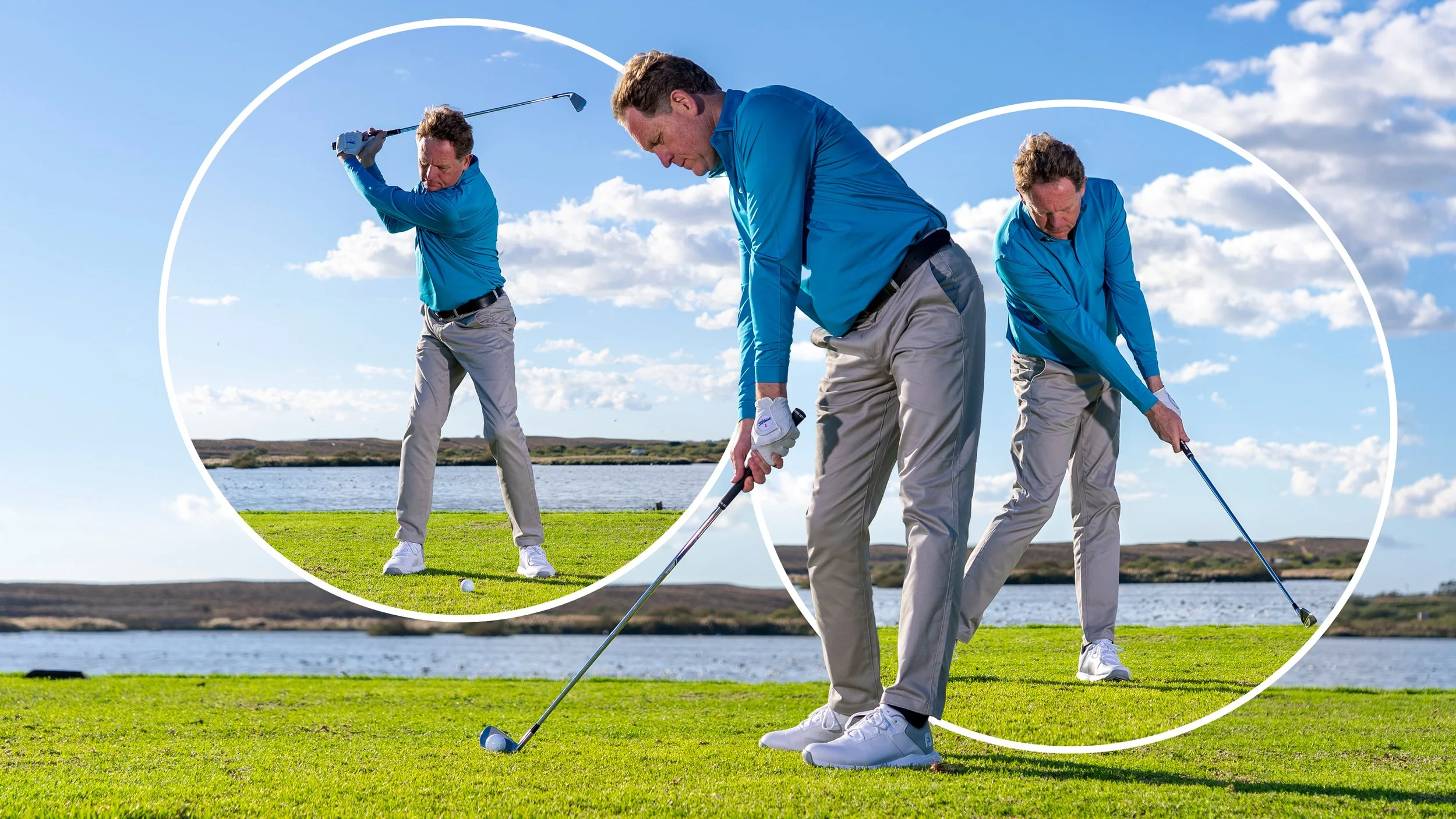
Why the Golf Swing Stresses the Knee
Although golf may appear less physically demanding than cutting or contact sports, the golf swing (depending on the club used) can generate high rotational, deceleration, and ground-reaction forces that challenge the ACL and surrounding structures.
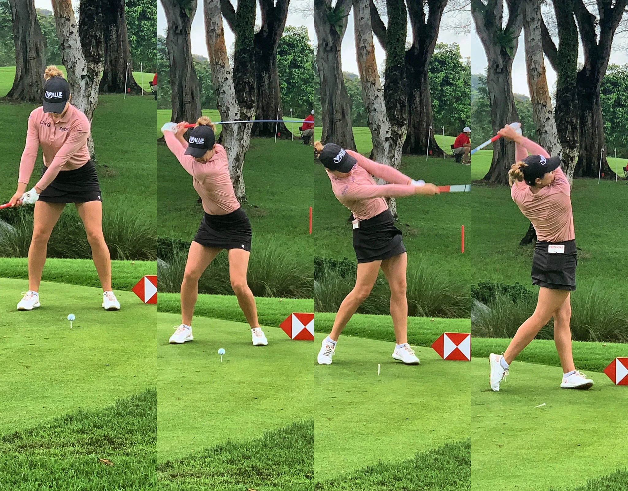
Rapid rotation and weight transfer: During the downswing, the hips and knees must rotate in a coordinated sequence while maintaining balance and trunk stability.
Ground-reaction forces: Both lead and trail legs experience significant vertical and horizontal loads, particularly at impact and follow-through.
Asymmetrical loading: The lead and trail legs are exposed to different force profiles and understanding these is essential to prevent reinjury.
Research shows that:
The lead knee typically experiences higher external knee moments (valgus/adduction) compared to the trail leg (Lynn et al., 2023).
Vertical ground reaction forces at the lead knee are often larger during impact and follow-through, increasing stress on the ACL (Castro et al. (2018).
Peak knee adduction and rotational moments challenge soft tissue structures, reinforcing the importance of targeted rehabilitation and load management (Baker et al., 2017).
These biomechanical demands explain why some golfers struggle with knee discomfort or compensatory swing mechanics even months after ACLR.
Lead vs Trail Leg: Implications for Golfers
Lead Leg (Target Side) ACLR
Role in the swing:
The lead leg acts as the primary braking and weight acceptance limb during follow-through. It stabilises the body and absorbs deceleration forces as the golfer rotates through impact.
Rehabilitation and RTS Implications:
Emphasise eccentric control, frontal-plane stability, and rotational strength
Introduce controlled deceleration and lateral load drills before progressing to full swings
Prioritise dynamic balance and weight-transfer practice, particularly on the lead leg
Lead-leg ACLR often presents a challenge for golfers because the knee must tolerate high external moments under rotational load, and improper rehabilitation can increase risk of reinjury or swing compensation.
Trail Leg (Back Foot) ACLR
Role in the swing:
The trail leg contributes primarily to power generation, initiating rotation and push-off during the backswing and downswing. Although it experiences lower vertical loads than the lead leg, the trail knee is critical for torque production and rotational sequencing.
Rehabilitation and RTS Implications:
Quadriceps and hamstring strength symmetry ≥90%
Balanced single-leg hops, deceleration drills, and lateral load tolerance
Gradual increase in rotational drill speed and swing intensity
Symptom-guided practice sessions; no pain or swelling
Golf-Specific Progression:
Chipping and pitching practice for fine motor control
Half-speed swings with short irons
Full-speed swings with long irons and drivers
Gradual progression from short course rounds to full 18-hole play
Key Principles:
Progress based on objective performance, not time post-surgery
Include rotational control and frontal-plane stability in all drills
Maintain symptom-free practice with careful monitoring of fatigue
Assessing Readiness for Golf
A combination of objective and subjective criteria provides the best assessment of readiness:
Objective Measures:
Quadriceps and hamstring strength ≥90% limb symmetry
Hop test battery ≥90% symmetry
Controlled deceleration and rotation drills
Movement Quality:
No dynamic valgus on squats or lunges
Stable pelvis and trunk during rotational activities
Smooth weight transfer between lead and trail legs
Psychological Readiness:
Confidence with swing volume and speed
Low fear of reinjury
Positive scores on tools such as ACL-RSI or IKDC
Optional Advanced Testing:
Swing video analysis
Force-plate assessment for weight transfer and rotation
Fatigue-based stability drills to simulate 18-hole play
Summary
Returning to golf after ACL reconstruction is more than a timeline — it’s about restoring function, strength, and confidence in a sport that places unique rotational and deceleration demands on the knee. Lead and trail legs serve distinct biomechanical roles, meaning that rehabilitation and return-to-play strategies must be leg-specific. Psychological factors such as fear of reinjury or lack of confidence are among the strongest predictors of delayed return to sport or altered movement patterns
A safe return requires:
Objective functional assessments
Progressive, performance-based load management
Attention to rotational control, frontal-plane stability, and hip/trunk sequencing
When approached systematically, golfers can return to play stronger, more resilient, and with reduced risk of reinjury.
References
Alarifi, S. M., Herrington, L. C., Althomali, O. W., et al. (2025). Persistent biomechanical alterations at clearance to return to sport indicate that athletes may not be fully prepared for safe loading. Orthopaedic Journal of Sports Medicine. 13(5)
Baker, M. L., et al. (2017). Biomechanical factors leading to high loading in the anterior cruciate ligament of the lead knee during the golf swing. Sports Medicine, 47(8), 1657–1669.
Castro, M. A., Fernandes, O., Silva, L., Marta, S., Vaz, J., Cabri, J., & Pezarat‑Correia, P. (2018). Lead and trail legs ground reaction forces and timing during the golf swing with different clubs in average golfers. Advances in Research on Foot and Ankle, ARFA‑109.
Lynn, S. K., Wang, J., Schmitt, A. C., & Barnes, C. L. (2023). Lower body joint moments during the golf swing in older adults: comparison to other activities of daily living. JSSM, 22(3), 382–388.
Nawasreh, Z., Logerstedt, D., Cummer, K., et al. (2018). Functional performance at 6 months predicts return to pre-injury activity at 12 and 24 months following ACL reconstruction. British Journal of Sports Medicine, 52(6), 375–382.
Wright, A., Reid, D., & Potts, G. (2025). Return to sport (RTS) tests and criteria following anterior cruciate ligament reconstruction: A scoping review. Knee Journal. Knee. 2025 57:179-199